Blog
By Jennifer Webster
While widespread use of mammograms has reduced mortality from breast cancer, specialists at St. Luke’s University Health Network have developed an algorithm to help providers offer additional, nuanced screenings that take into account individual variations in breast density. At the same time, St. Luke’s University Health Network is incorporating advanced technology to aid early detection.
Many area physicians have received a letter from Joseph P. Russo, MD, Section Chief of Women’s Imaging at St. Luke’s University Health Network, introducing St. Luke’s algorithm for Individualized Breast Screening. The letter and the program it heralds offer guidance regarding a new Pennsylvania law that requires mammography providers to notify women if they have dense breast tissue and inform them that dense breasts may be more prone to cancer and make detection more difficult. The letter also provides clarity to physicians about screening recommendations.
“Historically, we have had few options to deal with screening challenges related to dense breasts,” Dr. Russo says. “However, with the increasing popularity of screening modalities such as MRI, ultrasound and 3-D imaging, we have more tools available.”
Expert Guidance
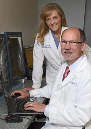
Breast density is broken down into four categories, from least to most dense. “There is some variability in how radiologists interpret breast density on a mammogram,” notes Tricia A. Kelly, MD, breast surgical oncologist at St. Luke’s University Health Network. “In general, however, density expresses the percent of fibroglandular elements in the breast.”
Women who are notified that they have dense breasts are asking their providers what they should do, in addition to mammography, to detect breast cancer early, Dr. Russo says. Providers, in turn, are drawing on St. Luke’s University Health Network’s expertise.
“Primary care physicians and gynecologists in the community are looking to us for help in triaging these patients,” he says. “We have developed our algorithm to guide clinicians and patients.”
The Individualized Breast Screening algorithm has its basis in a breast-screening decision tool released by the California Breast Density Information Group (CBDIG) in 2013. Like CBDIG’s algorithm, it draws on the Tyrer-Cuzick risk model for breast cancer. In mammogram reports created by St. Luke’s University Health Network, patients’ Tyrer-Cuzick profile appears, in addition to breast density information.
Risk Management
Breast density is not the only or primary indication a woman is at heightened risk for cancer. Risk profiles include family history, especially when there are multiple cases of breast or related cancers — such as ovarian and pancreatic — at early ages. Prior biopsies with atypical findings, use of hormone replacement therapy, and not having children before age 30 or having no children also increase breast cancer risk, Dr. Kelly says.
A small population of men is at risk, as well, she adds. “Men who are asymptomatic for breast cancer but who carry the BRCA gene are recommended to undergo screening mammography,” Dr. Kelly says. “Most cancers are sporadic, not genetic, but of the small percent of inherited cancers, if we identify a gene mutation in a patient — typically a woman — then we might suggest male and female family members be screened.”
St. Luke’s University Health Network tailors the most appropriate screening regimen to each patient’s individual risk factors.
“Many screening tests involve radiation exposure, and all increase the risk of unnecessary biopsy,” says Lee B. Riley, MD, PhD, Medical Director of Oncology at St. Luke’s University Health Network. “We balance those side effects with the hope of an early diagnosis.”
Systematic Approach
“Personalized medicine has been used in many disciplines. However, the concept hasn’t historically been applied to breast screening. At St. Luke’s, we’re changing that. We’re tailoring screening based on risk factors, including breast density, to develop an individualized screening program. We anticipate this will result in increased and early detection of cancer.”
— Joseph P. Russo, MD, Section Chief of Women’s Imaging, St. Luke’s University Health Network
Standard mammogram remains the gold standard for breast screening, Dr. Russo says, and that is reflected throughout St. Luke’s algorithm. Other screening modalities are adjuncts, not substitutes, to mammography, providing vital information for patients with certain risk factors.
Women who have average to intermediate risk plus dense breasts may benefit from automated breast ultrasound (ABUS), a refinement of a technique for characterizing different lesions — distinguishing a cyst from a solid nodule, for example. In this procedure, available at St. Luke’s University Health Network, the sonographer places a compression plate on top of the breast then activates an ultrasound transducer that scans automatically, resulting in six series of images. Imaging the breast in a unified way, this technology acquires images independently of the skill of the sonographer, making the results more reproducible than they have been in the past. Importantly, no radiation is required.
“ABUS picks up subtle findings anywhere between the nipple and the chest wall,” Dr. Kelly says. “That makes it an excellent modality to look at breast tissue in a different way.”
Another well-established screening modality, breast MRI is recommended for women at high risk for breast cancer, regardless of their breast density. Available at St. Luke’s University Health Network, this sensitive screening approach is most appropriate for women who have a heightened index of suspicion about breast cancer due to family history or other risk factors, as it balances an increased number of false positives with an excellent rate of cancer detection.
Finally, 3-D tomosynthesis, a method for obtaining 3-D X-ray images of the breast in very small slices, is expected to be available at St. Luke’s University Health Network in the near future. Three-D tomosynthesis is suggested for women who are at intermediate or high risk for breast cancer and have dense breast tissue.
“St. Luke’s anticipates acquiring the latest generation of 3-D mammography, which promises significant advantages over the existing forms of tomosynthesis in terms of quality and radiation exposure,” Dr. Russo says.
“St. Luke’s has a philosophy of offering multiple modalities and providing a clear plan detailing when to use each,” he adds. “This approach differentiates us from the majority of health systems.”
Prioritizing Early Detection
Early detection of breast cancer makes survival more likely and treatments less invasive. “As a surgeon, I have more options when cancer is detected at an earlier stage,” Dr. Kelly says. “If the cancer is smaller, I can offer a patient breast conservation, whereas with a large tumor, I would advise a mastectomy.”
“If we discover the cancer early, it may not have metastasized,” Dr. Riley adds. “Patients whose cancer has spread do not fare as well, whereas patients whose cancer is detected in the ducts have a 98 percent 10-year survival rate.”
Benefits of an Extensive Network
St. Luke’s University Health Network uses a hub-and-spoke model to provide mammography to patients from throughout the Lehigh Valley. Nine sites perform standard screening mammograms, while a central location — housing a team of experienced technologists, radiologists and breast health nurses under one roof — handles follow-up and diagnostic breast screenings. Patients referred for additional screening have the opportunity to consult directly with a radiologist, according to Dr. Riley.
“Each patient has unique factors,” he says. “Our algorithm is tailored to guide the referring provider in making sure all the relevant factors have been discussed. The patient, referring physician and radiologist work together to arrive at a decision.”
“Physicians need to be aware that there are multiple indications for breast screening,” Dr. Kelly says. “At St. Luke’s University Health Network, our specialists are happy to advise referring physicians and, if needed, see their patients for an evaluation.”
Making the Case
The screening algorithm developed by St. Luke’s University Health Network may benefit patients seeking insurance coverage for screenings in addition to mammography.
“Insurance companies typically favor detecting disease earlier, when it is less expensive to treat,” notes Lee B. Riley, MD, PhD, Medical Director of Oncology at St. Luke’s University Health Network. “This algorithm helps physicians make the case for a particular screening when they are arguing on a patient’s behalf.”
To learn more, visit www.sluhn.org/cancer or call 866-STLUKES.